
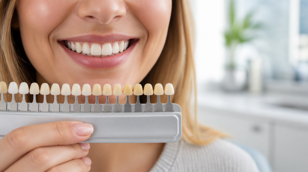
How Whitening Results Differ on Canine vs Incisor Teeth
Why Canine Teeth Whitening Works Differently Than You Expect Canine teeth whitening is one of the most misunderstood steps in any smile-brightening routine — and the confusion starts before you even open the gel. Your canines were never the same shade as your front teeth. Clinical shade evaluations confirm a definitive baseline difference, with maxillary … Read more
Why Canine Teeth Whitening Works Differently Than You Expect
Canine teeth whitening is one of the most misunderstood steps in any smile-brightening routine — and the confusion starts before you even open the gel. Your canines were never the same shade as your front teeth. Clinical shade evaluations confirm a definitive baseline difference, with maxillary canines measuring visibly darker than central incisors even in healthy, untreated smiles. This is not a hygiene failure. It is structural anatomy.
Canines carry a heavier concentration of pigment from the moment they erupt. Positioned at the corners of the arch and built to handle the mechanical stress of tearing and lateral chewing, they evolved with greater dental mass and higher chromatic saturation. That extra saturation is precisely what makes them resist bleaching agents that move incisors several shades in a matter of days.
When a patient looks in the mirror after two weeks of whitening and notices the canines still look warm or “yellowy,” they are usually seeing a pre-existing shade gradient that the treatment made more visible — not one it created. The incisors leapt ahead because they were designed to. Understanding that distinction is the foundation of every realistic whitening plan.
The Enamel-to-Dentin Ratio That Sets Each Tooth’s Whitening Ceiling
Every tooth has a whitening ceiling — a maximum lightness achievable before additional gel exposure adds risk without adding brightness. That ceiling is governed by the ratio of enamel thickness to dentin bulk, and canines and incisors sit at opposite ends of that spectrum.
Tooth color is dominated by dentin, the yellow-brown core beneath the enamel. Enamel is semi-translucent, so the thinner it is, the more dentin shows through — but enamel is also the medium through which peroxide diffuses to reach and oxidize pigment molecules inside. Incisors have thinner enamel and comparatively less dentin bulk, meaning bleaching agents penetrate faster and encounter a smaller reservoir of color to break down.
Canines flip this equation entirely. They are denser, with a thicker dentin core and more saturated pigment throughout the entire tooth structure. Peroxide must travel through more tissue and oxidize a larger volume of color molecules before the shade shifts even one step. Clinical research confirms incisors reach chromatic saturation — their whitening ceiling — significantly faster than canines, with canines requiring more sessions or longer protocols to achieve comparable lightness.
Sensitivity patterns follow the same logic. Because incisors have thinner enamel, peroxide diffuses toward the pulp more quickly, producing sharper sensitivity during in-office treatment. Thinner enamel is a double-edged structure: faster color response, greater nerve irritation. No gel concentration overrides this anatomy. A canine will always require more time, and forcing a higher-strength product does not raise its ceiling — it raises your risk of sensitivity and gum damage.
How Tooth Shape Affects Gel Contact and Coverage
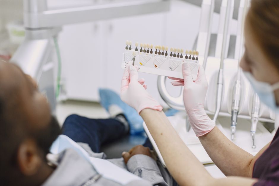
Anatomy shapes not just how peroxide penetrates, but how reliably it stays in contact with the tooth surface during treatment. Incisors present a broad, flat, forward-facing plane — an ideal surface for a strip or tray reservoir to sit flush against for the entire application window.
Canines are curved and pointed. Their rounded labial surface and prominent cusp tip cause flat whitening strips to tent over the peak and lift away at the edges, reducing sustained gel contact exactly where the thicker enamel body needs it most. Whitening progresses fastest at the incisal and gingival margins where enamel is naturally thinnest, and slowest across the dense central body of the tooth. On a pointed canine, that central body is proportionally larger, so coverage gaps there translate directly into stubborn residual color.

This geometry is the primary reason custom-fabricated trays consistently outperform one-size strip formats for even results. A tray molded to the patient’s arch wraps the canine’s curvature and holds gel against the full labial surface. Generic strips and boil-and-bite trays cannot conform with the same precision.
A familiar scenario: a patient completes a standard strip course and achieves bright, even incisors alongside noticeably shadowed canines, then concludes the product failed. In reality it worked on every surface it genuinely contacted. The solution is not a stronger formula — it is sustained, conforming coverage over the curved teeth.
Why Incisors Peak Weeks Before Canine Teeth Whitening Is Complete
Timing is where the canine-versus-incisor gap becomes most disorienting, and it is the single most common driver of premature product abandonment. The two tooth types do not simply whiten to different degrees — they whiten on entirely different schedules.
Controlled clinical research tracking color change over time found that incisor shade stabilized at roughly six weeks, while canine color continued shifting and only stabilized around twelve weeks — essentially double the timeline under the same treatment protocol. During that middle window, the incisors have reached their peak while the canines are still actively responding. This is precisely when most people quit, convinced the product has plateaued when the canines are, in fact, still catching up.
Studies examining in-office protocols using high-concentration hydrogen peroxide document the same pattern: canines require a greater number of sessions to harmonize with the incisors already whitened in earlier appointments. The disparity is not a product deficiency — it is a predictable biological timeline that needs to be scheduled around, not fought against.
There is a meaningful bright side to that lag. Long-term comparative research found that once treatment continued to completion, canines showed a significantly stronger increase in lightness than incisors, ultimately producing more homogeneous lightness values across the arch. The tooth that starts slowest can close much of the gap — but only if the protocol runs its full course.
Delivery method influences this timeline meaningfully. Comparative research found that in-home bleaching produced greater color changes in canines than in-office treatment, while causing less sensitivity overall. The extended wear time and lower peroxide concentration of home trays appears to suit the canine’s need for prolonged, sustained exposure — a slow drip rather than a surge.
Managing the Two-Tone Smile Effect After Canine Teeth Whitening
The two-tone smile — bright incisors flanked by noticeably warmer canines — is the most common cosmetic complaint following short, aggressive whitening courses. It develops when treatment stops at the moment the incisors peak but before the canines have had sufficient time to catch up.
The critical first point: in most cases this effect is temporary and self-correcting if treatment continues at a sensible cadence. Because canines keep responding for weeks longer, extending the protocol gives them room to close the gap organically. Stopping abruptly is what locks the two-tone appearance in place.
The second point is that a modest gradient is not only normal but can read as natural. Since canines start darker at baseline, a finished smile where they remain a touch warmer than the central incisors often looks authentic rather than artificial. The single-shade, uniformly bright result is an aesthetic preference, not a biological default — and chasing it with escalating gel strength carries real cost in sensitivity and tissue irritation.
For a persistent gradient that continues beyond the expected canine catch-up window, targeted touch-ups are far more effective than retreating the entire arch. Some professional systems allow gel to be applied selectively to lagging canines via syringe and custom tray, delivering additional sessions where needed without over-treating already-whitened incisors. This is a considerably safer strategy than increasing concentration across every tooth.
A scenario worth recognizing early: completing a strip course immediately before a major event, noticing the two-tone effect three days out, and reaching for a higher-strength emergency product. The smarter approach is finishing whitening at least a month ahead of the date and reserving the final weeks for canine-only supplemental sessions. Planning ahead is the only reliable way to control the timing gap.
Practical Strategies for Even Results Across All Tooth Types
Even results come from working with tooth anatomy, not overriding it. The following approaches consistently narrow the canine-incisor gap without escalating risk.
- Choose conforming delivery over flat strips. Custom or well-fitted trays wrap the curved canine surface and maintain gel contact where strips tent away. If strips are your only option, press them firmly around each canine and reseat any section that lifts during the session.
- Plan for the longer canine timeline from day one. Expect incisors to peak in weeks and canines to need roughly double that window. Schedule whitening well ahead of any event so canines have time to reach a harmonious shade without last-minute pressure.
- Favor lower-concentration, longer-contact protocols for saturated canines. The extended wear time of home bleaching trays appears to suit canine anatomy better than brief, high-intensity in-office bursts, and comparative evidence links it to greater canine color change alongside lower sensitivity.
- Use selective touch-ups rather than blanket intensity increases. Apply extra sessions only to the lagging canines. Raising gel concentration across every tooth to force a faster canine response overexposes incisors and elevates sensitivity unnecessarily.
- Track each tooth against its own baseline. Photograph your smile under consistent lighting before treatment begins. Progress evaluated tooth-by-tooth against its starting shade prevents the false conclusion that a slow canine signals a failed product.
- Manage incisor sensitivity proactively. Because thinner-enamel incisors zing first, consistent use of a desensitizing toothpaste containing potassium nitrate or stannous fluoride before and during treatment helps you stay the course long enough for canines to catch up.
One clinically important exception: when discoloration stems from fluorosis rather than pigment saturation, the typical canine lag can disappear entirely. Research examining a microabrasion-plus-bleaching protocol for mild-to-moderate fluorosis found no statistically significant color difference between central incisors, lateral incisors, and canines at treatment completion. When the discoloration is surface-based and intrinsic rather than saturation-based, the usual canine-incisor hierarchy does not apply — a reminder that identifying the cause of discoloration shapes the entire protocol.
For a deeper look at how peroxide concentration affects tissue safety across different tooth types, the guide to teeth whitening safety and sensitivity covers the clinical thresholds that separate effective treatment from overexposure. And if you’re deciding between professional and over-the-counter options for your canine teeth whitening plan, the comparison of professional versus at-home whitening methods breaks down the tradeoffs in gel strength, tray fit, and overall outcome.
When Uneven Shades Mean You Should Stop and Consult a Dentist
Most canine-incisor unevenness is predictable physiology. But certain patterns signal conditions that surface whitening cannot resolve and may actively worsen — knowing the difference protects both your teeth and your time.
Seek professional evaluation if a single tooth appears dramatically darker than its neighbors — particularly a canine or incisor that looks grey or brown rather than simply less bright. A lone dark tooth frequently indicates internal changes: old trauma, a compromised pulp, or a failing restoration. These require diagnosis and targeted intervention before any bleaching begins.
White patches or opaque streaks appearing during treatment are another stop signal. That pattern can reflect underlying enamel defects or fluorosis that demand specific protocols — such as microabrasion combined with bleaching — rather than additional gel applications. More of the same product will not correct a structural issue.
Persistent, sharp sensitivity that does not settle between sessions warrants a pause and professional input. Because incisors diffuse peroxide rapidly through thin enamel, they can flag pulp irritation early. Pushing through severe sensitivity risks lasting nerve irritation and gum damage. A dentist can adjust concentration, reduce session length, or transition you to a gentler home protocol suited to your specific anatomy.
Whitening agents sold in professional settings are regulated for concentration and safety standards. Over-the-counter products occupy a different regulatory category, and the gap between them matters when sensitivity or unusual color responses appear. A dentist can assess whether the reaction you’re experiencing is expected physiology or a sign that the current approach needs to change.
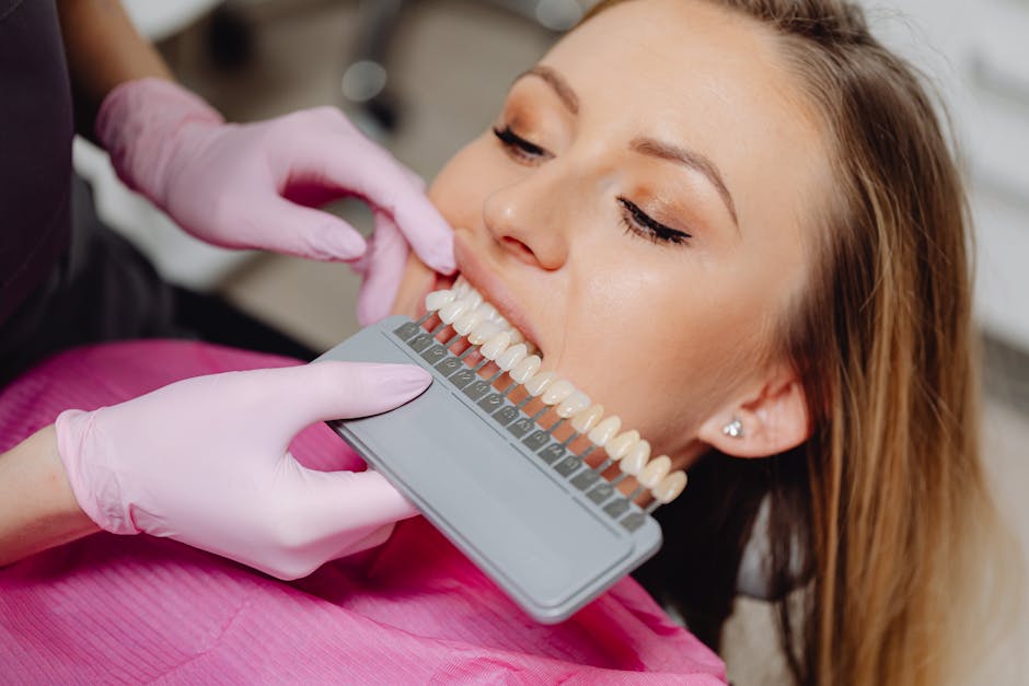
The bottom line: canine teeth whitening is a longer, more geometry-dependent process than whitening front teeth, but it is a manageable one when the protocol respects both the timeline and the anatomy. Most people who understand the canine lag from the start finish treatment satisfied. Most people who don’t, quit early and blame the product for a result that was still in progress.
References
- Clinical evaluation of different types of teeth in in-office whitening. PubMed, National Library of Medicine.
- Color matching between incisors and canines in dental bleaching protocols. Research, Society and Development Journal.
- Color differences of canines and incisors in a comparative long-term clinical trial. PubMed, National Library of Medicine.
- Controlled clinical trial addressing teeth whitening with hydrogen peroxide. Clinics, Elsevier.
- Comparison of efficacy and outcome satisfaction between in-office and home teeth bleaching. Journal of Oral Science, JSTAGE.
- A comparison of three whitening protocols for mild-to-moderate dental fluorosis. PMC, National Library of Medicine.
- Predictable tooth whitening outcomes: expert clinical analysis. Dentistry.co.uk.
Disclaimer
This article is for informational purposes only. LLRNCARE makes no representations or warranties about the completeness, accuracy, reliability of the information. Any reliance is at your own risk.
For professional dental advice, consult a qualified dental professional. For regulatory compliance, consult legal experts.